Nomi, Jason S.; Bzdok, Danilo; Li, Jingwei; Bolt, Taylor; Chang, Catie; Kornfeld, Salome; Goodman, Zachary T.; Yeo, B.T. Thomas; Spreng, R. Nathan; Uddin, Lucina Q. Imaging Neuroscience 2 (2024): 1-13. .╠¤
In brain imaging studies using╠¤resting-state fMRI, scientists often detect a signal called the╠¤global signal (GS). While this signal is known to include noise and artifacts (like breathing or head motion), it also carries important information about brain activity that reflects a personтАЩs mental state and traits.╠¤
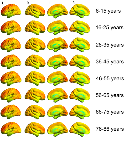
This study looked at how the GS changes with╠¤age, using data from people aged╠¤6 to 85. Researchers found that different brain regions contribute to the GS in different ways across the lifespan.╠¤
- Subcortical areas╠¤(deep parts of the brain), such as the╠¤thalamus╠¤and╠¤putamen, showed╠¤linear changes. The thalamus contributed more to the GS as people got older, while the putamen contributed more in younger people.╠¤
- Another deep brain structure, the╠¤nucleus basalis of Meynert, showed a╠¤U-shaped pattern: stronger contributions in children and older adults, and weaker in middle-aged adults. A similar pattern was seen in╠¤cortical regions╠¤(outer brain areas), especially in networks related to attention and thinking (like the╠¤frontoparietal network).╠¤
These patterns suggest that the GS is made up of two layers: one from subcortical areas and another from cortical areas, and that their roles in the GS change with age.╠¤
Overall, this research shows that the GS isnтАЩt just noiseтАФit contains meaningful brain activity that╠¤changes throughout life. Because of this, scientists should be cautious when choosing to remove it in studies that explore how the brain develops or ages.╠¤
╠¤
Fig. 1.╠¤
Average global signal topography across 10-year age groups. Increased associations between the GS with visual, sensorimotor, and prefrontal cortical regions are found across each age group.╠¤